Cardiac CT
Overview
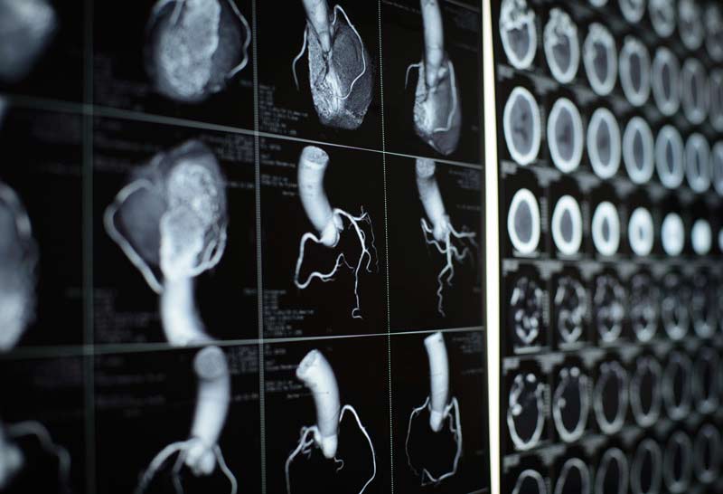
At Indotaj Medical Center in Tajikistan, a cardiac computed tomography (CT) scan is a sophisticated procedure employing multiple X-ray beams at various angles to capture precise, three-dimensional (3D) images of the heart, great vessels, and adjacent structures.
Our state-of-the-art cardiac CT technology, with the option of intravenous (IV) contrast (dye), enhances the visualization of cardiac structures and blood vessels. Utilizing multi-slice scanning, our healthcare professionals obtain high-resolution, dynamic 3D images, providing detailed insights into the anatomy and function of the heart and great vessels.
Why it's done
Coronary Artery Disease (CAD) Assessment:
- Cardiac CT is often used to evaluate the coronary arteries, detecting the presence of plaque, stenosis, or other abnormalities that may indicate coronary artery disease.
Calcium Scoring:
- Calcium scoring, a subset of cardiac CT, assesses the amount of calcium deposits in the coronary arteries. Higher calcium scores may suggest a higher risk of heart disease.
Evaluation of Coronary Anomalies:
- Cardiac CT helps identify any congenital or acquired anomalies in the coronary arteries, providing valuable information for treatment planning.
Assessment of Cardiac Chambers and Valves:
- The procedure allows for detailed imaging of the heart chambers and valves, helping diagnose conditions such as valve stenosis, regurgitation, or structural abnormalities.
Evaluation of Cardiac Tumors or Masses:
- Cardiac CT can detect and characterize tumors, masses, or other abnormalities within the heart, aiding in the diagnosis and treatment planning.
Aortic Disease Assessment:
- It is used to assess the aorta for conditions like aneurysms, dissections, or other abnormalities that may impact vascular health.
Preoperative Planning:
- Before certain cardiac surgeries or interventions, a Cardiac CT may be performed to provide detailed anatomical information for preoperative planning.
Assessment of Cardiac Function:
- In some cases, Cardiac CT can provide information about the overall function of the heart, including the ejection fraction and cardiac output.
Evaluation of Pulmonary Veins:
- Cardiac CT is used to assess the pulmonary veins, especially in the context of conditions like atrial fibrillation or prior to certain cardiac procedures.
Follow-Up after Cardiac Interventions:
- Patients who have undergone cardiac interventions such as stent placement or coronary artery bypass surgery may have follow-up Cardiac CT scans to assess treatment outcomes.
Risk
- Radiation Exposure
- Contrast Dye Allergy
- Kidney Issues with Contrast Dye
- Risk for Pregnant Women
- Contrast-Induced Nephropathy
- Rare Complications
How do I get ready for coronary artery bypass surgery?
Jewelry and Attire:
- Kindly leave your jewelry at home. Upon arrival, you’ll change into a hospital gown to facilitate the imaging process.
Heart Rate Management:
- If necessary, your healthcare provider may administer medication to temporarily slow down your heart rate, optimizing the quality of the images obtained.
Contrast Administration:
- If contrast (dye) is required for the procedure, a skilled nurse will insert an intravenous (IV) line into a vein in your arm. The contrast enhances the visibility of blood vessels and the heart on the scans.
Positioning on Scanner Table:
- You will lie on your back on a specialized table designed to move into the central space of our advanced round CT scanner.
Electrode Placement:
- Three small areas on your chest will be cleaned, and adhesive electrode patches will be applied. These electrodes connect to an electrocardiogram (EKG) monitor, recording your heart’s electrical activity during the examination. Any chest hair that interferes with electrode placement may be gently shaved.
Body Positioning:
- While lying on the scanner table, you’ll raise your arms above your head. Your head and feet will extend beyond the scanner, ensuring a comprehensive view without compromising your comfort.